Start with the Question, Not the Magnification
The best microscopy method is the one that reveals the evidence needed to answer your question. Tracking how cells move, locating a protein, examining a fractured material, and resolving a membrane require different capabilities. Before choosing an instrument, define the smallest feature you must distinguish, whether the specimen must remain alive, and what kind of contrast will identify the feature.
Magnification alone is a poor guide. Enlarging an image does not recover details the instrument failed to resolve. Resolution describes the ability to distinguish nearby features, while contrast determines whether those features stand out from their surroundings. Field of view, imaging speed, and the number of samples you need to examine also matter. A highly detailed image of a tiny region may be less useful than a broader, repeatable measurement.
Resolution: What Each Method Can Reveal
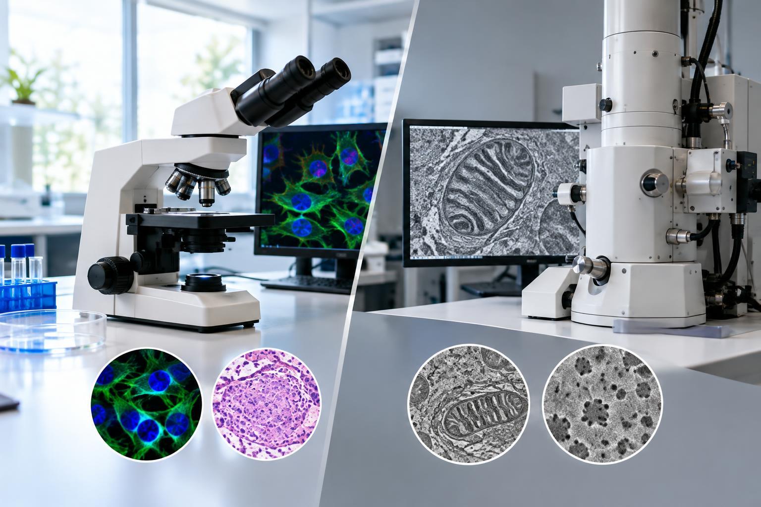
Conventional light microscopy typically resolves lateral details separated by roughly 200–300 nanometers under favorable conditions. Resolution along the depth axis is generally poorer. This makes light microscopy useful for cells, tissues, and many larger subcellular structures, but not for reliably separating every closely spaced molecular feature. Confocal imaging improves optical sectioning and reduces out-of-focus blur; it does not automatically provide nanoscale resolution.
Super-resolution light microscopy can reveal finer details, although performance depends on the technique, labeling, specimen, and acquisition conditions. Electron microscopy can resolve much smaller features because electrons have far shorter wavelengths than visible light. However, useful resolution is not determined by wavelength alone: electron optics, specimen thickness, preparation, stability, and radiation damage all affect the result.
Choose a resolution target tied to a measurement. If you only need cell counts or tissue organization, electron microscopy may add complexity without improving the answer.
Sample Compatibility and Preparation
Light microscopy is usually the starting point for observing living systems. Depending on the method, samples can remain in liquid, under controlled temperature, and within an environment compatible with growth. Brightfield and phase-contrast imaging can examine some specimens without fluorescent labels. Fluorescence adds specificity, but labeling may alter biology, and illumination can cause photobleaching or phototoxicity.
Electron microscopy generally requires conditions incompatible with living specimens, including a vacuum and substantial preparation or freezing. Conventional biological preparation may involve fixation, dehydration, embedding, sectioning, or conductive coatings, depending on the imaging mode. These steps can introduce shrinkage, extraction, or other artifacts. Materials samples may also need careful cutting, polishing, or thinning.
Cryogenic electron microscopy preserves specimens in a frozen state and can avoid some artifacts associated with drying or chemical fixation. It does not show living activity, and successful vitrification, specimen thickness, and beam sensitivity remain important constraints.
Choose the Imaging Mode That Matches the Evidence
Within light microscopy, fluorescence is especially valuable when the question concerns the location or behavior of a labeled molecule. Widefield imaging can support efficient surveys and time-lapse experiments. Confocal microscopy helps separate structures at different depths, while light-sheet approaches can be useful for volumetric imaging with reduced out-of-plane illumination. The right choice depends on sample size, transparency, speed, and acceptable light exposure.
Electron microscopy also encompasses distinct tools. Scanning electron microscopy, or SEM, is commonly used to examine surface morphology and related contrast. Transmission electron microscopy, or TEM, uses electrons transmitted through thin specimens to reveal internal ultrastructure. Both have specialized modes that extend their capabilities, including compositional or crystallographic analysis.
Structural detail does not automatically establish molecular identity. Electron-dense features may require additional labeling or complementary measurements for interpretation. Conversely, a fluorescent signal can identify a target without resolving its surrounding ultrastructure.
Balance Detail Against Throughput and Reliability
Instrument access, operator expertise, preparation time, and analysis requirements can determine whether an experiment is practical. Electron microscopy often involves specialized facilities and demanding workflows. Advanced optical methods can also require considerable expertise, particularly when reconstruction, drift correction, or quantitative fluorescence measurements are involved.
Consider how many independent samples and regions you need, not just how impressive one image could look. Very small fields of view can make representative sampling difficult. High-resolution three-dimensional datasets can also create substantial storage and analysis burdens.
Build controls into the imaging plan. Check whether fixation changes morphology, whether labels are specific, and whether image processing introduces misleading features. Keep acquisition settings consistent where comparisons require it, and distinguish observations supported by raw data from interpretations dependent on reconstruction or segmentation.
Use Complementary Methods When Necessary
A practical decision rule is to start with light microscopy for live behavior, labeled molecular localization, or broad surveys. Favor SEM when surface structure is central, and TEM when fine internal structure is essential. Consider super-resolution optical methods when finer spatial detail and fluorescent specificity must be combined.
When no single method answers the question, correlative light and electron microscopy can connect a labeled region with its ultrastructure. This requires compatible preparation and careful image registration. A small pilot study is often the best next step: test whether the proposed workflow produces interpretable, repeatable evidence before committing to a larger experiment.